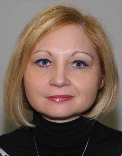
European Society of Radiology: Sports imaging is the main theme of IDoR 2019. In most countries, this is not a specialty in itself, but a focus within musculoskeletal radiology. In your country, is there a special focus on sports imaging within radiology training or special courses for interested radiologists?
Vladka Salapura: Sports imaging is predominantly included within the field of musculoskeletal radiology in Slovenia, as it is in most European countries. During the training, our residents gain basic knowledge and initial experience in this type of pathology as they receive six months of training in MSK radiology in university educational facilities. Special courses and lectures are organised by the Society of Musculoskeletal Radiology, which is a part of our national Association of Radiology, for all interested radiologists, and they are generally very well attended.
ESR: Please describe your regular working environment (hospital, private practice). Does sports-related imaging take up all, most, or only part of your regular work schedule?
VS: The Clinical Institute of Radiology is part of our largest University Hospital, University Medical Centre Ljubljana, Slovenia. We employ 55 radiologists, and are currently training 76 residents. We perform around 360,000 imaging studies and minimally invasive treatments per year. Our work is organised at the level of subspecialty work as we perform mostly diagnostic and therapeutic examinations dedicated to a specific organ system. We have nine radiologists dedicated to the MSK field. We are closely connected to trauma surgeons and orthopaedic surgeons in our daily practice. As a result, our work is closely connected to acute and chronic sports injuries and their late consequences for at least one third of our working time.
ESR: Based on your experience, which sports produce the most injuries that require medical imaging? Have you seen any changes in this regard during your career? What areas/types of injuries provide the greatest challenge to radiologists?
VS: The most prominent injuries are those with direct contact or significant forces between the athletes (boxing, wrestling, hockey, rugby, netball), and sports that include high velocities or quick reactions with sudden changes of motion such as skiing or football. In our practice, we see a lot of skiing injuries as this is a very popular sport in the area. The greatest challenges for radiologists include rare injuries of the small intra-articular structures, evaluation after multiple repeated injuries, or assessment after an operative treatment, as these findings are harder to interpret.
ESR: Please give a detailed overview of the sports injuries with which you are most familiar and their respective modalities.
VS: The sports injuries I most often see in my clinical practice are sports injuries of the knee, shoulder, and ankle joints. Knee injuries are present in at least half of the patients that come to our department. These include injuries of anterior cruciate ligament and menisci, cartilage lesions, collateral ligaments injuries, as well as posterior cruciate ligament injuries and posterior-lateral corner injuries, which are rarer. The best modality for intra-articular types of injuries is magnetic resonance imaging of the knee. Very often these injuries also include bone fractures, which may require a computed tomography for complete evaluation before surgical treatment. Soft tissue injuries and injuries of extra-articular structures may be initially evaluated with ultrasound.
In the shoulder joint, the main problem is shoulder instability after luxation. The modality that gives us the most detailed information is magnetic resonance arthrography, which we perform routinely under ultrasound guidance. Most often we find classic labral or bone Bankart lesions of the anterior-inferior labral complex and Hill-Sachs lesions of the posterior superior part of the humeral head. Seldom, shoulder luxation can be posterior, in which case we find ‘reverse Bankart lesion’ signs. SLAP (superior labral tear from anterior to posterior) lesions of the superior labrum are also common in sports with overhead throwing motions (baseball, volleyball, American football, swimming or tennis). These injuries are also most accurately diagnosed with magnetic resonance of the shoulder with intra-articular contrast injection. Lesions of the rotator cuff (athletes, heavy lifting) are also common and cause pain with subsequent subdeltoid bursal effusion and motion limitations, especially arm lifting. In extra-articular lesions isolated only to the soft tissues, we initially perform ultrasound of the shoulder.
In the ankle joint, the most common injuries arise in the ligaments. The complex anatomy of the ankle joint is demanding for imaging and evaluation. The most proper modality is magnetic resonance of the ankle, which offers the possibility to show the whole extent of the injury including small intra-articular ligaments, cartilage and subchondral bone defects. The evaluations of milder injuries limited to the superficial extra-articular areas are adequately shown with ultrasound.
ESR: What diseases associated with sporting activity can be detected with imaging? Can you provide examples?
VS: Diseases associated with sporting activity can be acute or chronic. In acute events we mostly deal with fresh injuries of joints and muscles. These include joints as well as hand and wrist injuries which often occur in boxing and weightlifting. Depending on the mechanism and the force of the injury, these can include bone and ligament or tendon injuries, especially complex injuries of the knee, or the so-called ‘terrible triad’. To diagnose these injuries, it is crucial to be aware of the mechanism of the injury and to be aware of all clinical data available. It may be necessary to use different radiology modalities in complex cases and the choice of imaging modality very much depends on the suspected clinical injury. In chronic injuries we mostly deal with overuse injuries affecting lower limb (Achilles and Patella tendinopathy, plantar fasciosis, stress injuries of the foot and ankle) or upper limb such as tendinopathy of common extensor tendons in the elbow or common flexor tendon, aka tennis elbow and golfer’s elbow respectively. Complex or intra-articular injuries usually demand imaging with magnetic resonance. When dealing with soft tissue extra-articular injuries, ultrasound is a very helpful modality. Muscle injuries most often include hamstring and calf muscles.
ESR: Radiologists are part of a team; for sports imaging this likely consists of surgeons, orthopaedists, cardiologists and/or neurologists. How would you define the role of the radiologist within this team and how would you describe the cooperation between radiologists, surgeons, and other physicians?
VS: Cooperation between clinicians involved in the diagnostics and treatment of sports injuries and radiologists is of crucial importance for the successful treatment of our patients. Modern medicine does not exist without collaboration between different medical fields. The role of the radiologist is very important during the whole diagnostic and treatment process as they should be closely involved with clinicians. We should be present at interdisciplinary meetings and available for advice and discussion. The clinicians, however, should provide radiologists with specific clinical questions and available clinical findings to make the evaluation of the radiologist more meaningful and competent.
ESR: The role of the radiologist in determining diagnoses with sports imaging is obvious; how much involvement is there regarding treatment and follow-up?
VS: During the treatment and follow-up, the role of the radiologist is equally important. The question that is often asked after the injury is how soon an individual can return to a normal activity and start training or competing again. To be objective and more accurate in answering this question, the control and follow-up examinations are performed. It is of crucial importance to compare the control findings with previous examinations as well as evaluation of control findings. The work of a radiologist is finished only when the rehabilitation of the patient is successfully completed.
ESR: Radiology is effective in identifying and treating sports-related injuries and diseases, but can it also be used to pre-empt them? Can the information provided by medical imaging be used to enhance the performance of athletes?
VS: Radiology is effective in identifying sports-related injuries and new techniques that are quickly developing provide us with even more accurate tools for the depiction of early changes or possible changes that may become important in the future. As much as these advances are important for medical science and may help in the preservation of anatomical structures, we must take into consideration that this is only a single aspect of anticipated events. The human body is very complex, and it would be naïve to make determinations based only on advanced imaging findings without taking all of the other numerous factors into consideration.
ESR: Many elite sports centres use cutting-edge medical imaging equipment and attract talented radiologists to operate it. Are you involved with such centres? How can the knowledge acquired in this setting be used to benefit all patients?
VS: Most elite sports centres are aware of the fact that only high-quality work and the combination of excellent clinical and research work can bring long-term prosperity along with a stable, positive environment that is motivational for young, talented people. We can all benefit from their experience, published research and educational work. These centres have resources to conduct large, multicentre, high-quality studies and the results of their work may influence the clinical practice of other centres in this field. It is crucial that leading sports centres give importance to this aspect of their work and use their technical and human resources for educational and research work as well.
ESR: The demand for imaging studies has been rising steadily over the past decades, placing strain on healthcare budgets. Has the demand also increased in sports medicine? What can be done to better justify imaging requests and make the most of available resources?
VS: This is a big question in almost every country today. The number of imaging studies is growing, and it seems we still do not have a definitive solution for this. Better assessing the appropriateness and justification for radiological procedures are key elements in this issue. The ESR iGuide platform is one very important systemic solution for this problem. If we manage to implement this clinical decision support system worldwide, we may be able to limit the growing number of imaging studies as evident in some countries thus far. We should also be aware of the quality of the studies performed. Maintaining high standards and quality of imaging studies (regarding protocols, technical aspects and reporting) will prevent unnecessary additional studies and consequently lower the cost of diagnostic processes. This should be of interest to all included parties; radiologists, national healthcare systems and insurance companies.
ESR: Athletes are more prone to injuries that require medical imaging. How much greater is their risk of developing diseases related to frequent exposure to radiation and what can be done to limit the negative impacts from overexposure?
VS: We must follow the ALARA (As Low as Reasonably Achievable) principles for every radiological procedure. Protection from radiation during the procedures is nowadays very good and modern equipment allows for limited exposure to professionals and patients during procedures. If we follow the instructions and stick to the rules applied to radiation protection, we will be well protected even if diagnostic studies are repeated. There is no scientific data to support the notion of there being a greater risk of developing diseases related to radiation exposure in athletes.
ESR: Do you actively practise sports yourself and if yes, does this help you in your daily work as MSK radiologist?
VS: I practise recreational swimming and I enjoy it very much. My favourite style is crawl. I think swimming is one of the best ways to practise sports and stay in shape while minimising the possibility of sports injuries! Previously, I tried skiing for a couple of years and suffered a complex knee injury. I do find it helpful that I can connect to the problems of our patients on an informal level as well. We generally tend to understand others better when we go through a similar experience ourselves.
 Vladka Salapura, PhD, MD is Associate Professor of Radiology and Vice-Chairman of the Institute of Radiology at University Hospital Ljubljana. She is Head of the Musculoskeletal Department at her Institution. She has held the position of Vice-President of the Slovenian Association of Radiology since 2015, and she is President of the National Advisory Board of Radiology at the Ministry of Health. She has been a permanent member of the Cathedra of Radiology at the Medical University Ljubljana since 2011. As an active researcher, she is an expert in musculoskeletal and interventional radiology. Her main field of expertise includes minimally invasive treatment of the musculoskeletal system. She has co-authored more than 30 peer-reviewed papers, 115 conference abstracts and ten book chapters, and has won several international awards.