European Society of Radiology: Sports imaging is the main theme of IDoR 2019. In most countries, this is not a specialty in itself, but a focus within musculoskeletal radiology. In your country, is there a special focus on sports imaging within radiology training or special courses for interested radiologists?
Tomás Pascual: Yes, every year, there are specific musculoskeletal radiology courses, which focus on sports injuries and their management using magnetic resonance imaging (MRI) and ultrasound. Our team members always participate in these kinds of activities, either as speakers or attendees. At the same time, every year, our team offers a musculoskeletal radiology fellowship that lasts two years. This position involves assisting others and fulfilling academic tasks, such as participating in conferences, courses, congresses, and publishing scientific papers in journals.
ESR: Please describe your regular working environment (hospital, private practice). Does sports-related imaging take up all, most, or only part of your regular work schedule?
TP: I work in two private diagnostic imaging centres dedicated specifically to sports and trauma injuries in both amateur and professional athletes. In our country, most of our patients are athletes who play football, rugby, tennis or hockey. We usually work together with orthopaedic surgeons and kinesiologists, using all imaging techniques.
ESR: Based on your experience, which sports produce the most injuries that require medical imaging? Have you seen any changes in this regard during your career? What areas/types of injuries provide the greatest challenge to radiologists?
TP: I think rugby is the sport that requires diagnostic imaging the most. This has to do with the fact that rugby practice triggers constant trauma and demands, physical exercise. In my opinion, muscle injuries are still a major challenge for radiologists, given their incidence rate and difficult interpretation. Throughout the years, the study and classification of these kinds of injuries have been perfected, allowing players to get back in the game faster and reduce both the recurrence and relapse rates.
ESR: Please give a detailed overview of the sports injuries with which you are most familiar and their respective modalities.
TP: The injuries we see the most in our daily practice are muscle and tendon injuries. There is also a high prevalence of athletic pubalgia. We always evaluate these cases using ultrasound first and only in specific situations do we use MRI; depending on the location and size of the injury, affected muscle, etc. Other injuries we see frequently are traumatic ones; mostly in knees, ankles, and shoulders, particularly in football and rugby players. In these cases, we apply MRI in the first place, especially when we believe there are ligament or intra-articular injuries. We use computed tomography (CT) when we think there might be a fracture. In the case of chronic intra-articular injuries, such as shoulder labral tears and chondral injuries due to instability or microinstability or femoroacetabular impingements in the hip, MR and CT arthrography is the most specific and sensitive method.
ESR: What diseases associated with sporting activity can be detected with imaging? Can you provide examples?
TP: With ultrasound, we see many injuries related to athletic pubalgia, such as sports hernias and adductor tendinopathies. These occur in sports where there are aggressive rotation movements in the hip while running, as in football, rugby and tennis. We also find a high prevalence of Achilles and hamstring tendinopathies in runners and patellar tendon injuries in basketball and volleyball players. There is a high rate of overuse injuries such as stress fractures in tibiae, hips and ankles in runners, and particularly in patients who run recreationally but under high physical demand. Trauma injuries such as fractures, shoulder dislocations and knee or ankle sprains are very frequent in contact sports.
ESR: Radiologists are part of a team; for sports imaging this likely consists of surgeons, orthopaedists, cardiologists and/or neurologists. How would you define the role of the radiologist within this team, and how would you describe the cooperation between radiologists, surgeons, and other physicians?
TP: The role of radiologists in these teams is fundamental both for diagnosing the pathology and for monitoring its progress and response to treatment. We even have the possibility of participating in the treatment of some injuries through minimally invasive procedures. Fluent contact with our colleagues is utterly important in this chain of work. This continuous exchange of opinions and information allows a prompt and adequate diagnosis, which then leads to the best treatment option.
ESR: The role of the radiologist in determining diagnoses with sports imaging is obvious; how much involvement is there regarding treatment and follow-up?
TP: Radiologists are becoming more actively involved in many sports and are now a part of the medical team together with orthopaedists and kinesiologists. In our day-to-day practice, we play a major role in the follow-up and monitoring of injuries to get the players back in the game and back to their usual physical activity quickly.
ESR: Radiology is effective in identifying and treating sports-related injuries and diseases, but can it also be used to prevent them? Can the information provided by medical imaging be used to enhance the performance of athletes?
TP: Some imaging techniques can prevent or reduce the incidence of injuries in certain sports. For instance, in our country, we request a routine lateral x-ray of the cervical spine in all rugby players to detect if there is spinal stenosis and prevent spinal cord injuries. We also perform ultrasound screenings in professional rugby and football teams before they start long competitions. We systematically study all the muscle groups of the lower limbs, looking for acute, sub-acute and chronic muscle injuries or scars. We evaluate the midsection of the body to detect weak spots in the abdominal wall, and we study the adductor, patellar and Achilles tendons for detecting tendinopathies or after-effects. Then, according to the findings, we can plan a preventive or therapeutic kinesic treatment.
ESR: Many elite sports centres use cutting-edge medical imaging equipment and attract talented radiologists to operate it. Are you involved with such centres? How can the knowledge acquired in this setting be used to benefit all patients?
TP: We work in two high-performance sports centres where we have state-of-the-art high-field MRI, CT and ultrasound scanners. Besides having the necessary equipment, I think it is essential to have adequate knowledge of the most prevalent pathologies and their mechanisms. Working with orthopaedists and kinesiologists helps us detect which aspects of the images are more relevant for reaching a prompt and proper treatment.
ESR: The demand for imaging studies has been rising steadily over the past decades, placing strain on healthcare budgets. Has the demand also increased in sports medicine? What can be done to better justify imaging requests and make the most of available resources?
TP: Yes, the demand for imaging studies in sports has risen considerably over the last few decades, and this has to do with several factors. To begin with, there has been a decrease in the cost of the studies that, mainly due to an increase in supply, which allows better access not only for professional athletes but also for amateurs. At the same time, orthopaedists and sports physicians rely more on our specialisation to avoid making mistakes in terms of diagnosis and therapy. Imaging techniques allow them to detect the injury and its stage, and to monitor its progress. To justify ordering these studies, it is important to communicate the main indications, advantages and disadvantages of each technique. Working as a team gives us the opportunity to treat the patient as a whole, emphasising the importance of clinical signs and symptoms to determine the right technique for confirming the presumptive diagnosis.
ESR: Athletes are more prone to injuries that require medical imaging. How much greater is their risk of developing diseases related to frequent exposure to radiation and what can be done to limit the negative impacts from overexposure?
TP: It is important not to order tests unnecessarily to avoid overexposure, more so if the clinical signs and symptoms do not justify such scans. As an alternative, we can use ultrasound or MRI scans if these are useful for the kind of pathology being treated. New digital tomography and radiology machines operate with very low doses of radiation and provide high-quality images. This makes them a great alternative to avoid the harmful effects of overexposure.
ESR: Do you actively practise sports yourself and if yes, does this help you in your daily work as MSK radiologist?
TP: I play football and practise CrossFit and kitesurf on a daily basis. The first two have helped me understand and interpret important aspects of the injuries that may occur and their mechanics, especially in the case of trauma and overuse injuries, which are so typical in these sports.
European Society of Radiology: Sports imaging also applies to sports-related injuries of the brain. In case you are familiar with this, please also answer the following questions:
ESR: Which sports have the highest risk of inducing brain injuries?
TP: Contact sports such as rugby and boxing.
ESR: What imaging modalities do you use with traumatic brain injury specifically in athletes?
SN: We use both computed tomography and magnetic resonance imaging.
ESR: What can be learned from sports-related injuries that can be applied to a broader use, for example, those sustained through automobile or other accidents that cause traumatic brain injury?
TP: Knowing the clinical factors and understanding the mechanism of injuries can contribute to detecting the scope of injuries that may occur and therefore, it can help us select the right protocols for detecting said injuries.
ESR: How have advances in brain imaging allowed you to predict patient outcomes more accurately?
TP: The improvements in image quality, the reduction of times and the development of new MRI sequences all allow earlier detection of injuries and a better characterisation and detection of their stage, aspects which are fundamental for the diagnosis and prognosis.
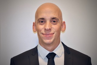
 Dr. Tomás Pascual is coordinator in the ultrasound service of HIMAN Imaging Centre and part of the medical staff of IMAXE Diagnostic Centre in Buenos Aires, Argentina. He serves as a member of the Argentine Society of Radiology (Sociedad Argentina de Radiología, SAR), where he collaborates in the musculoskeletal division. His area of specialisation is diagnostic imaging applied to sports and trauma injuries, particularly in high performance athletes. Dr. Pascual has authored and co-authored several papers, which were presented at both national and international congresses such as the ECR, RSNA and CADI (Argentine Congress of Diagnostic Imaging). He has participated as a lecturer in many courses and conferences in his home country and abroad.
His most important contributions deal with athletic pubalgia, Achilles tendinopathy and the study of muscle injuries in athletes.