European Society of Radiology: Could you please give a detailed overview of when and for which diseases you use cardiac imaging?
Narumol Chaosuwannakit: Non-invasive cardiac imaging procedures provide essential information for the detection, diagnosis, and management of cardiovascular diseases, and have a vital role in risk assessment and clinical decision-making. The range of diagnostic tools used to evaluate cardiovascular diseases have expanded over the past decade; in particular computed tomography (CT) and magnetic resonance (MR) have emerged as viable alternatives to echocardiography, exercise electrocardiography (ECG), and invasive angiography.
Guidelines for the appropriate use of each imaging modality developed in the United States and Europe are often not applicable in Asian countries because of the differences in the healthcare systems, medical expenses, body habitus, and disease prevalence between Asian and Western countries.
For this reason, the Asian Society of Cardiovascular Imaging (ASCI) separately developed ASCI appropriate use criteria (AUC) for cardiac CT and MR in 2010. The top ten clinical indications for advanced cardiac imaging are summarised as follows:
- Symptomatic: non-acute chest pain suspected of being stable coronary artery disease with an uninterpretable ECG and/or unable to exercise
- Symptomatic: acute chest pain suspected of being an acute coronary syndrome with a normal ECG or equivocal cardiac biomarker
- For evaluating the patency of coronary artery bypass graft (CABG) or percutaneous coronary intervention with stent implantation (PCI, left main stent)
- Newly developed or suspected heart failure for the evaluation of ischaemic or non-ischaemic ethology
- Cardiac evaluation prior to non-coronary cardiac surgery or vascular surgery
- Anomalous coronary artery disease
- Kawasaki disease without a previous definitive test available or for a follow-up
- Equivocal prior test; such as exercise stress test ECG or nuclear imaging; such as SPECT or PET
- Congenital heart disease both pre-operative and post-operative
- Cardiac mass, pericardial disease and great vessels
ESR: Which modalities are usually used for what purpose?
NC: Cardiovascular magnetic resonance (CMR) is now a mature imaging modality and has become the gold standard technique for evaluating the myocardial function, quantifying myocardial volumes, and detecting myocardial scars. CMR also has the unique ability to provide detailed tissue characterisation, including the assessment of oedema, iron overload, and diffuse myocardial fibrosis.
In addition, MRI has a key advantage over most other imaging modalities when it comes to imaging the heart. That advantage is the ability to differentiate between various kinds of tissue based on multiple and different tissue characteristics. For instance, with CT and x-ray imaging, we rely on density: the denser the tissue, the whiter it appears; the less dense it is, the darker it is. If tissue densities are not very different, then we would be unable to distinguish them on a CT or x-ray scan unless we administer contrast.
Furthermore, with ultrasound or echocardiography (ECG), we use the ability of the tissue to propagate sound to generate images. MRI provides us with many different ways to look at tissue, such as T1- or T2-weighted images, fat-suppressed imaging, perfusion imaging, diffusion imaging, and so on. There is even MRI spectroscopy that looks at the biochemical signature of different tissues.
With MRI, the range of tools plus the technological advances that enable to expedite entire processes give us much more diagnostic power. We use cardiac MRI for the evaluation of myocardial scars or fibrosis, various types of cardiomyopathy, myocardial viability, tissue characterisation of cardiac tumours, and a comprehensive evaluation of the cardiac function.
Coronary/cardiac CTA is mainly used for the evaluation of the cardiac morphology in various clinical scenarios, as mentioned above.
ESR: What is the role of the radiologist within the ‘heart team’? How would you describe the cooperation between radiologists, cardiologists, and other physicians?
NC: The politics of cardiac imaging vary from place to place. In some centres, radiologists operate cardiac MRI; in others, the cardiologists carry out all the imaging. At our institution, we have a cardiology fellow in our department who works with radiology residents to perform cardiac imaging studies. As such, an interdisciplinary approach is rewarding for everyone, especially the patient.
One of the methods that radiologists can use to facilitate the appropriate allocation of resources is clinician education. Performing the appropriate exam can save the patient both money and allows them to avoid ionising radiation associated with unnecessary/unindicated exams.
Additionally, considering the rapid technological advances in radiology, regular clinico-radiographic meetings and lectures could greatly enhance the clinicians’ ability to order the appropriate studies. While the majority of studies ordered by clinicians are appropriate, occasionally the radiologist may feel that there is a more suitable investigation for the clinical question. Communicating this concern to the ordering clinician can result in the collaborative selection of the appropriate study and a learning opportunity for both parties. This type of collaboration between radiologists and clinicians can be taken a step further by jointly developing clinical decision rules or guidelines for imaging.
ESR: Radiographers/radiological technologists are also part of the team. When and how do you interact with them?
NC: I personally discuss the protocols for each cardiac imaging study that is tailored for each patient with the radiographer for the best quality images. Some nurses are also engaged in the radiology department, and they carry out the more care-related tasks, especially in cardiovascular radiology.
In clinical practice, radiologists and radiographers are the ones who conduct cardiac imaging. The radiologist is responsible for the entire imaging examination, from its prescription to the delivery. The radiologist has to justify the chosen imaging modality, define the data acquisition procedure, interpret images according to the medical history and the status of the patient, and provide clear and reliable documents that answer the questions raised by physicians.
Moreover, the role of the radiographer has grown with the increasing complexity of our techniques. The radiographer has to ensure the patient’s safety prior to and during the examination, to conduct and adapt the imaging procedures according to the patient’s condition and the findings of the examination, to post-process images; to producing and sending images to a picture archiving and communicating system (PACS). Most of these procedures are defined according to standardised protocols.
ESR: Please describe your regular working environment (hospital, private practice). Does cardiac imaging take up all, most, or only part of your regular work schedule? How many radiologists are dedicated to cardiac imaging in your team?
NC: I work at a tertiary care heart centre and medical school hospital at the Faculty of Medicine. Cardiac imaging takes up about 80% of my regular work schedule. Unfortunately, I am the only radiologist who is specialised in cardiac imaging in our department.
ESR: Do you have direct contact with patients and if yes, what is the nature of that contact?
NC: Yes, I do have contact with patients. I examine their history and conduct a further physical exam in case we do not have enough clinical data, and I explain the procedure and/or respiratory training before scanning.
ESR: If you had the means: what would you change in education, training and daily practice in cardiac imaging?
NC: The traditional view of a radiologist in a darkroom in front of a viewing box adding value to the healthcare system solely by interpreting images is rapidly becoming out-dated. While advances in imaging technology, especially cardiac CT and cardiac MRI, have expanded the radiologist’s diagnostic capabilities, the value of radiologists has also expanded outside of image interpretation.
Advances in information technology have the potential to make the role of the radiologist more central in the overall systems operation of the healthcare system; specifically by providing more immediate clinically relevant information, enhancing patient safety, and improving public health. In addition, radiologists can perform a gatekeeper role by significantly reducing imaging costs resulting from the ordering of inappropriate and/or self-referred studies.
ESR: What are the most recent advances in cardiac imaging and what significance do they have for improving healthcare?
NC: I would say the most recent advance in cardiac MRI is the non-contrast parametric mapping technique. Given recent safety concerns of using gadolinium in patients with impaired renal function, there has been renewed interest in the development of techniques, which can perform tissue characterisation without the need for contrast agents.
These techniques rely on the intrinsic magnetic T1, T2, and T2* relaxation properties of the myocardium. By measuring images with different sensitivities to these intrinsic properties, parametric maps can be created. Native T1 mapping is acquired before the administration of exogenous agents to provide a quantitative map of the T1 relaxation times of the tissues being imaged on a pixel-by-pixel basis. Increased native T1 is useful for detecting acute myocardial pathologies; such as oedema, infarction, myocarditis, and sub-acute cardiomyopathies – such as cardiac amyloidosis, hypertrophic cardiomyopathy, and dilated cardiomyopathy – as well as for assessing diffuse fibrosis.
Low global myocardial T1 values have clinical utility in a myocardial iron overload (siderosis) and Fabry disease. Native T1 mapping can also detect increases in the myocardial blood volume associated with the coronary vasodilation downstream of significant coronary stenosis. This may potentially be an emerging application to detect myocardial ischaemia without the need for the administration of gadolinium.
In addition to its ability to diagnose disease, there is increasing evidence that native T1 mapping carries prognostic power in risk stratification including in patients with acute myocardial infarction, amyloidosis, and dilated cardiomyopathy. Novel acquisition strategies combined with advanced reconstruction techniques are enabling high-resolution, true 3D dynamic acquisitions, which will be the future of CMR. Recent developments of fast T1, T2, and T2* mapping techniques are rapidly entering the clinical CMR arsenal of techniques for tissue characterisation.
The recent advance in cardiac/coronary CTA would be spectral CT, which has proven particularly helpful in vascular applications, with the ability to quantify arterial plaque in contrast-enhanced vessels. In a traditional black-and-white CT image, it can be difficult, if not impossible, to differentiate between the iodine and calcium in the artery; spectral imaging can display the materials as two distinct colours and the iodine contrast can be fine-tuned on the spot. Spectral CT can provide information which conventional CT could not give us and we can also minimise the contrast to as low as 20 ml and then use post-processing software to improve diagnostic performance and make the diagnostic CTA picture clearer than conventional CT.
ESR: In what ways has the specialty changed since you started? And where do you see the most important developments in the next ten years?
NC: The past decade has witnessed significant improvements in cardiovascular imaging, which is all to the benefit of better diagnosis, management, and early prevention of cardiovascular disease. Going forward, I expect the observed growth and refinements in imaging technology and applications to continue unabated, from the high-end equipment of CT, CMR, 3D echocardiography and molecular imaging to miniaturisation with hand-held devices.
The future promises an unprecedentedly wide spectrum of opportunities. Early detection of disease and assessment of the cardiac phenotype in the early stages is paramount in preventing cardiovascular disease and particularly relevant in inherited diseases, where genetic markers are not yet available and conventional risk factors are absent.
There is also the possibility of using imaging for novel drug development and as a surrogate to the patient’s outcome, where appropriate.
Imaging will always be an integral part of clinical cardiovascular medicine. With new realities in healthcare emphasising quality and cost-effectiveness, future technologies will need to demonstrate value through greater efficiency and efficacy of care and/or patient outcomes.
Furthermore, greater emphasis will be placed on the appropriate utilisation of technology and resources including imaging. It is therefore imperative to avoid layering of multiple tests in individual patients; thus, we need to address both the cost and safety in the context of patient-centred care. Ultimately, we need to identify, through research, the best approaches to disease detection and management with a focus on providing the best care to the patient.
Spectral CT is rapidly replacing the traditional imaging system for several healthcare applications. As such, the highly advanced technology in spectral CT is expected to revolutionise the diagnosis and treatment procedures for various diseases in the foreseeable future. Thus, the technology is being increasingly used for the diagnosis of several diseases of the whole body as well as cardiac diseases.
Spectral CT is a budding technology, which is currently at its nascent stage. As per the latest report published by Market Research Future (MRFR), the global market for spectral CT is set to soar at a compound annual growth rate of 7.8% between 2017 and 2023.
ESR: Is artificial intelligence already having an impact on cardiac imaging and how do you see that developing in the future?
NC: Fully automated processing and high-level computer interpretation of cardiac imaging are becoming a reality. Application of machine learning to the vast amounts of quantitative data generated per scan and integration with clinical data also facilitates a move to more patient-specific interpretation.
These developments are unlikely to replace radiologists but will provide us with highly accurate tools to detect disease, stratify risk, and optimise patient-specific treatment. However, with each technological advance, we move further away from human dependence and closer to fully automated machine interpretation. There is a lot of hype and plenty of fear around artificial intelligence (AI) and its impact on the future of healthcare. As deep learning algorithms and narrow AI started to buzz, especially around the field of medical imaging, many radiologists went into panic mode.
However, let me compare the situation to that of the autopilot in aviation. The innovation did not replace real pilots; it augmented their tasks. On very long flights, it is handy to turn on the autopilot, but it is useless when you need to make rapid judgments. As such, the combination of humans and machines is the winning solution, and it will be the same in healthcare.
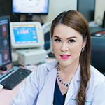
 Dr. Narumol Chaosuwannakit has been an Associate Professor of Radiology at the Faculty of Medicine, Khon Kaen University, and Division Head of Cardiovascular Imaging, Department of Radiology, and the Queen Sirikit Heart Centre of the Northeast, Khon Kaen, Thailand, since 2008.
She trained in cardiovascular imaging at Wake Forest University, North Carolina, USA, and specialises in advanced cardiovascular imaging including cardiac MRI and coronary/cardiac CTA.
Dr. Chaosuwannakit is currently one of the first fellows of the Society for Cardiovascular Magnetic Resonance (FSCMR) group. She has also regularly participated in several trials involving non-invasive methods for the diagnosis and treatment of heart diseases, such as tropical endomyocardial fibrosis, thalassaemia and systemic sclerosis.
Dr. Chaosuwannakit is a renowned national and international speaker with more than 50 presentations at major medical symposia to underscore the promise of cardiac CT or MRI as a comprehensive technique for assessing the heart and coronary arteries. She has authored or co-authored more than 40 papers, five books and 32 book chapters. Notably, she was the lead editor of a cardiovascular radiology book (in Thai).
Dr. Chaosuwannakit is a member of the Society of Cardiovascular Computed Tomography (SCCT), Society of Cardiovascular Magnetic Resonance (SCMR), European Society of Radiology (ESR), European Society of Cardiology (ESC), North American Society for Cardiovascular Imaging (NASCI), and Asian Society of Cardiovascular Imaging (ASCI). She is a former public relations committee member of the Thai Society of Cardiac Imaging (TSCI).