European Society of Radiology: Could you please give a detailed overview of when and for which diseases you use cardiac imaging?
Marco Francone: Modern cardiac imaging virtually encompasses the whole spectrum of cardiovascular pathologies, from congenital pathologies to ischaemic heart disease and cardiomyopathies. A recent, and still developing, field of interest regards the so-called setting of toxic cardiomyopathies, which includes quite a heterogeneous group of affections analysing effects of various ‘substances’ on the myocardium, from so-called recreational drugs to chemotherapy and chronic exposure to various pharmacological agents. A more specific subspecialty within cardiac imaging is the so-called ‘cardio-oncology’, analysing the complex interaction between new biological and antiblastic agents and myocyte oxidative metabolism.
ESR: Which modalities are usually used for what?
MF: Cardiac CT (CCT) is the ‘gatekeeper’ to selective coronary angiography in patients with suspected coronary arteries disease, given its almost 100% negative predictive value which reliably allows excluding atherosclerosis before the invasive procedure. Furthermore, it can be used to comprehensively assess ischaemic heart disease and combine anatomic information with functional data regarding stenoses haemodynamics, which can be obtained through the evaluation of CT-derived fractional flow reserve and myocardial perfusion.
The main benefit of cardiac MR (CMR) is, conversely, tissue characterisation that is used to evaluate myocardial damage in an expanding variety of clinical scenarios, from ischaemic heart disease to the heterogeneous constellations of congenital and non-ischaemic cardiomyopathies.
Besides tissue changes, CMR is also regarded as the reference tool for the assessment of biventricular function and flow-dynamics which have critical importance to drive clinical-decision making in daily practice. This has conferred high clinical and prognostic relevance to the technique, which has progressively changed clinical paradigms in many ways. The recent development of the so-called ‘relaxometry’ imaging with T1 and T2 mapping allowed to further expand tissue characterisation capabilities of the method which may be considered as a sort of in vivo histology.
The cardiac imaging toolbox also includes widely diffused diagnostic methods like echocardiography and nuclear medicine derived techniques like scintigraphy and PET, which have been largely used for years and still represent reference and first-line approaches in a large variety of clinical settings.
ESR: What is the role of the radiologist within the ‘heart team’? How would you describe the cooperation between radiologists, cardiologists, and other physicians?
MF: Inter-specialty cooperation is the foundation of our profession as clinical radiologists. This is obviously mandatory in a complex and multidisciplinary field like cardiac imaging.
We have a fruitful and long-standing cooperation (sometimes friendship) with our cardiologists and clinical referrals. This cooperation derives from the awareness that any mutual exchange of information allows us to improve the quality of the service provided to patients. This is certainly the main goal of the medical profession.
ESR: Radiographers/radiological technologists are also part of the team. When and how do you interact with them?
MF: In my vision, radiographers should be part of a cardiac imaging team.
Our interactions are continuous and routine, particularly in long and sometimes complex examinations like CMR studies which can often offer an almost ‘real-time’ optimisation of acquisition protocols.
I believe that the strategic importance of subspecialty training for radiographers in cardiac imaging is generally underestimated in the radiological community.
This leads to a remarkable variability on the effective expertise of technicians working in cardiac imaging, which is sometimes even more pronounced than for doctors.
This had obvious reflections on the diagnostic quality of the exams and may therefore impact our daily work. Dedicated CV imaging radiographers are necessary in my opinion.
ESR: Please describe your regular working environment (hospital, private practice). Does cardiac imaging take up all, most, or only part of your regular work schedule? How many radiologists are dedicated to cardiac imaging in your team?
MF: Cardiac imaging makes up approximately 70% of my time, this also includes oncological routine.
I work in the largest university hospital in Italy and we have three CMR and CCT dedicated sessions per week requiring the support of nine people including two staff members, one PhD student and six part-time dedicated residents. In general, our mission is to stimulate our residents (overall almost 100 colleagues) to become familiar with cardiac imaging, trying to send the message that it will part of the clinical radiological routine in the future.
ESR: Do you have direct contact with patients and if yes, what is the nature of that contact?
MF: We try to have direct contact with most of our patients, particularly in the preparation phase in which to explain the nature of the exams, importance of certain parameters (i.e. heart rate etc.) and expected results (including possible weaknesses of various tests).
This is done via weekly outpatient visits sessions which are also held to filter inappropriate requests from general practitioners.
We are prepared to explain our reports, something which is frequently required by our patients.
ESR: If you had the means: what would you change in education, training and daily practice in cardiac imaging?
MF: Cardiac imaging is still perceived as an ultra-specialised field of diagnostic imaging, somewhat external to the routine activity of a general radiologist.
This is in partial contradiction with the epidemiology of cardiac diseases and with the wide diffusion of various imaging methods which have now become part of the clinical routine in most clinical settings. There is, therefore, a significant educational gap in our specialty training which could be filled with dedicated educational tracks that were systematically planned during the residency programme.
ESR: What are the most recent advances in cardiac imaging and what significance do they have for improving healthcare?
MF: In cardiac CT, the most significant technological advance has been given by the dramatic reduction of radiation dose, which could be achieved by combining ultra-low Kv acquisition setting with highly iterative reconstructions algorithms. These allowed to reach an average patient’s dose which is now consistently in the range of 1 mSv, leading to an expansion in the utilisation of the method in an increasing number of individuals, with possible future application for dedicated screening programmes in selected patients’ cohorts.
Cardiac MR has moved to a multiparametric imaging approach, which offers comprehensive morpho-functional information in all clinical settings. My favourite recently developed technical advance in CMR is the introduction of mapping imaging, which allows analysing and quantitatively assessing diffuse subtle tissue changes. This may provide relevant information for diagnoses with possible prognostic reflections.
A further future process to follow will be the wide diffusion of artificial intelligence in cardiovascular imaging, which may further change our paradigms in various imaging applications.
ESR: In what ways has the specialty changed since you started? And where do you see the most important developments in the next ten years?
MF: When I started cardiac imaging in 2001, the whole field was generally regarded as a niche application of radiology, with limited pioneering applications and subsequent minor clinical value.
Parallel perception of clinicians was negligible, having been indicated with expressions like ‘funny toy’ or ‘no more than pretty pictures’ in various international internal medicine and cardiology journals published in those years.
Today, after almost 18 years, various imaging methods (and specifically CT and MR) have been added to virtually all clinical guidelines of the various national and international societies, being milestones of the routine diagnostic pathway and of clinical scenarios.
The main challenge for the future will be to use this enormous potential wisely, creating dedicated services in all imaging centres and balancing slots within the so-called CT and MR routine, in light of a further exponential growth in the next few years.
ESR: Is artificial intelligence already having an impact on cardiac imaging and how do you see that developing in the future?
MF: ‘On demand’ access to high-performance computing and large healthcare databases will certainly support and sustain our ability to achieve personalised medicine.
Combining AI, big data and massively parallel computing will make it possible to create a revolutionary way of practising evidence-based, personalised medicine in cardiac imaging, supporting the activities of radiologists.
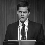
 Prof. Marco Francone is Professor of Radiology at the Department of Radiological, Oncological and Pathological Sciences at Sapienza University of Rome, Italy.
He is an internationally known expert in cardiac imaging and speaker at the most important international radiological and cardiological meetings including ECR, ESCR, ESOR, CIRSE, RSNA and the American College of Cardiology. His prominent field of research is cardiovascular imaging with both CT and MR, with a specific focus on myocardial infarction and cardiomyopathies. He is author and co-author of more than 100 indexed articles with an impact factor higher than 500.
Professor Francone is also Chairperson of the Scientific Committee of the European Society of Cardiovascular Radiology (ESCR) and was recently elected as President of the Italian College of Cardiac Radiology – Italian Society of Radiology.
He is also actively involved in the activities of the European Society of Radiology, having served as cardiac imaging subcommittee member for the European Congress of Radiology 2017–2019, and he was recently appointed as Scientific Subcommittee Chairperson for the 2020 meeting. He will also be a member of the ESR Research Committee until 2020.