Laura Dragonetti: Cardiac magnetic resonance (CMR) has made it possible to achieve great advances in cardiovascular diagnosis. The technique is non-invasive and does not use ionising radiation. A complete cardiological exam allows the evaluation of morphology, function and tissue characterisation (viability) all in just one study.
Another important instrument nowadays is coronary computed tomography angiography (CCTA), together with the development of volumetric equipment, has become an essential tool for diagnosing acute coronary syndrome in a non-invasive manner.
ESR: Which modalities are usually used for what?
LD: CCTA is used to evaluate coronary arteries, their anatomy (congenital or acquired anomalies), their course (visualising muscle bridges), or evaluating the coronary plaque in some degree of stenosis and calcification.
Regarding CMR, it is now the gold standard technique to evaluate ventricular volumes, myocardial function and mass. The additional capacity to characterise tissue makes it the ideal modality to use in clinical practice.
Image acquisition after contrast administration (Late Gad) and obtaining different enhancement patterns have contributed in the characterisation of typical injuries in a lot of pathologies, thus helping in the differential diagnosis of cardiomyopathies, by making a distinction between ischaemic and non-ischaemic disease.
Late Gad shows the damaged area of the myocardium, and thus can help the clinician decide on the location of a myocardial biopsy. It also offers risk stratification and prognostic value in many entities, such as hypertrophic cardiomyopathy, where the risk of arrhythmias and sudden death is always present.
Based on this, our group has been working on the study of different cardiomyopathies to reach an early diagnosis, carrying out studies on Fabry’s disease, amyloidosis, iron overload and recently incorporating Chagas’ disease.
ESR: What is the role of the radiologist within the ‘heart team’? How would you describe the cooperation between radiologists, cardiologists, and other physicians?
LD: The success of a procedure depends on good planning, and in this aspect, the role of the radiologist is fundamental and equally important to the rest of the team. Nevertheless, we feel that our work as radiologists is less considered.
It is clear that the election of a specific stent or valve number, as it usually occurs in TAVI procedures, are decisions that have to be taken as a group.
I believe, without a doubt, that it must be a cooperative decision made with the radiologist and the cardiologist, along with other specialists. And in our country, the relationship between both specialists might not be totally consolidated.
This is why our efforts in the Sociedad Argentina de Radiología (SAR) focus on achieving better cooperation between these two groups.
It’s true that a radiologist needs support from the cardiologist and the other way around, so both of them have to work together.
Good communication with other specialists is also important, for instance with haematologists, to value iron overload and its treatment with a totally safe method such as the T2*, or even with an oncologist to evaluate a ventricular dysfunction after treatment.
With this, I want to say that radiologists must increasingly participate in the creation of protocols and consensus between different specialties, and not only be spectators of how it is decided.
ESR: Radiographers/radiological technologists are also part of the team. When and how do you interact with them?
LD: The relationship between technologists and radiologists has always been a very good one. In the case of cardiac imaging, both technologists and doctors must be specifically trained to be able to work in this field.
It is important that they know the anatomy and the different study protocols based on the medical request of the patient. This is why it is also important to include the study motive and a brief review of the prescription.
Lately, I have noticed an increasing interest in performing cardiac studies, both in MRI and CT; these examinations will be requested more and more often in the years to come.
We have to be aware that nowadays conventional studies are not enough, and that modern radiology demands that technologists specialise in different areas, including cardiology, because their participation is fundamental.
Therefore it is necessary to move towards technologists’ capacitation and training so that we approach this new field without fear.
ESR: Please describe your regular working environment (hospital, private practice). Does cardiac imaging take up all, most, or only part of your regular work schedule? How many radiologists are dedicated to cardiac imaging in your team?
LD: I work at Alemán Hospital in Buenos Aires, a private community hospital, and other private centres – the High Technology Medical Institute (IMAT in Spanish), DIM, and Trinidad Palermo sanatorium. Most of these places have hospitalisation units, as well as outpatient care for cardiology. Cardiac imaging takes up most of my schedule.
Unfortunately, in our country, not many radiologists are dedicated to cardiac imaging. There are only a few of us, and my main role in the Education Committee of the SAR, which is led by Dr. Juan Mazzuco, was to include a cardiology module in the study plan of the specialist course, which doesn’t yet exist.
It is fundamental, from my point of view, that the radiologist knows the heart since it is not the patrimony of the cardiologist alone. This is why the imaging area must have specialists who know about the uses and benefits of CCTA and CMR.
The problem we face is that we need a high level of training and capacitation to start using these modalities.
Besides, it is usually necessary for young radiologists starting their career to train abroad, whether in Europe or North America, and this is very expensive. Moreover, there are very few possibilities for grants in our country.
The job opportunities are not enough, so finally, radiology trainees often decide for much faster job opportunities.
ESR: Do you have direct contact with patients and if yes, what is the nature of that contact?
LD: We do not have direct contact with the patients; they are referred by the cardiologist of the hospital or centre we work in.
ESR: If you had the means: what would you change in education, training and daily practice in cardiac imaging?
LD: Needless to say that having the means for education and training would be fundamental to develop cardiac imaging, and this is something I think of every day.
In our centres, we need to train a greater amount of doctors to learn and become interested in this subspecialty. We are now developing a project in that sense: the ‘Ultrafast Study’. This protocol trains local clinicians using local infrastructure to perform faster, cheaper and easier cardiac MR scanning – a key test in cardiology.
The scan takes around 20 minutes (instead of 1 hour) and we have scanned more than 70 patients to demonstrate it. Also, we provided a training course and international exchange to deliver a sustainable service with lasting links, as a pathfinder by local champions for national rollout.
The project took place in collaboration with Imaging Barts Heart Centre led by Professor James Moon and a visiting team, including Dr. Anna Herrey, Dr. Harold Litt, Dr. Juliano Lara Fernandez and Dr. Katia Menacho Medina.
The idea was to demonstrate that we can accelerate study times in our developing countries, and that with training and coaching we can succeed in carrying out more studies in the same amount of time that we currently do while remaining equally effective. We continue working on this right now. You can visit our webpage and even consult us at www.rapidcmr.com
The enthusiasm of all the participants and the good repercussion fundamentally on young physicians makes us think that only when different specialties work together – radiologists and cardiologists together –, will we achieve better results that help develop a modality we are passionate about, and to which we dedicate most of our professional (and also free) time.
ESR: What are the most recent advances in cardiac imaging and what significance do they have for improving healthcare?
LD: Without a doubt, the new techniques of mapping, among others, enable to visualise disorders at an early stage, demonstrating that CMR can modify the course of disease when a correct diagnosis is made. The latest advances enable to evaluate myocardial disorders early on, for instance T1, T2 and T2* mapping.
Sometimes it is not even necessary to administer contrast (native T1), which is indicated for patients with renal insufficiency and can be performed in Fabry’s disease evaluating the accumulation of anomalous substances in the tissue.
ESR: In what ways has the specialty changed since you started? And where do you see the most important developments in the next ten years?
LD: There were many changes in radiology and they have always been associated with the evolution of the latest available technologies and analysis software; new tools and artificial intelligence will soon come into play.
ESR: Is artificial intelligence already having an impact on cardiac imaging and how do you see that developing in the future?
LD: The use of big data will have a very important impact on future healthcare. The main causes of cardiovascular disease are auricular fibrillation, acute coronary syndrome and cardiac insufficiency, which cause more than 3.9 million deaths every year. We can develop early and precise therapies for these patients according to their risk profile, and bring different treatments to a good end.
Artificial intelligence makes medicine more specific, doctors and algorithms can evaluate similarly in some areas, but computers are faster.
But today our priority is the training of future generations of radiologists in all disciplines, including the heart, hoping they can improve the health of people.
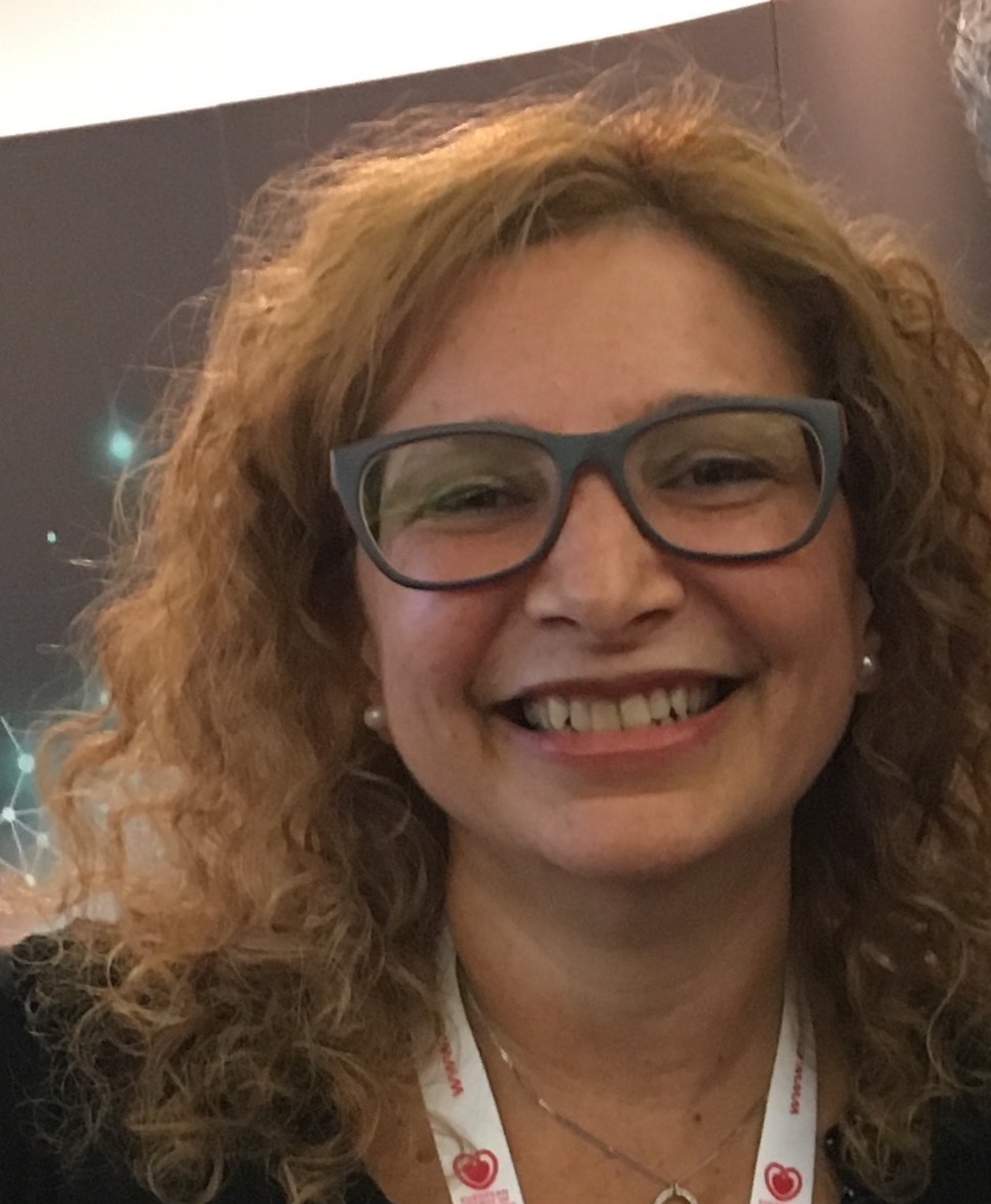
 Dra. Laura Dragonetti is an internal medicine doctor and cardiac radiologist; she is Head of the Cardiac Radiology Department at Alemán Hospital in Buenos Aires, Argentina.
She trained in general radiology in Buenos Aires, and in cardiac radiology in London, UK (Royal Brompton Hospital); Valencia, Spain (ERESA); and Brazil (INCOR). Since 1999 she has been an active researcher in multimodality cardiac radiology, with a special interest in MRI.
Dra. Dragonetti is an active speaker in Latin America, where she has been invited to give presentations. She is the author of many papers and more than 50 scientific posters and oral presentations.
She was director of the first International Course in Cardiac MR in Buenos Aires in June 2018. She served as a member of the Cardiac Imaging Committee of the Sociedad Argentina de Radiología (SAR) and is a current member of the SAR Education Committee. She is also a member of the Board of Cardiovascular Imaging of the Sociedad Argentina de Cardiología (SAC).
Dra. Dragonetti is a reviewer for the Argentinean Journal of Radiology (RAR) and a member of the European Society of Radiology, SCMR (Society of Cardiovascular Magnetic Resonance), Society of Cardiac Computed Tomography, European Society of Cardiology and European Association of Cardiovascular Imaging.